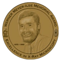
About the Award
The Werner Meyer-Ilse Memorial Award is given to young scientists for exceptional contributions to the advancement of X-ray microscopy through either outstanding technical developments or applications, as evidenced by their presentation at the International Conference on X-ray Microscopy and by supporting publications.
Nominees are qualified if they have performed this work as part of a completed Ph.D. thesis during the two-year period prior to and including the conference, or are expected to receive their degree in the very near future. The topics should be appropriate to the themes of the conference, and the work must be available to the award committee as conference papers, publications, or preprints at the time of nomination. Nominees must have submitted an abstract on their work requesting an oral presentation at the conference. The call for abstracts deadline of the conference is also the deadline for WMI award nominations.
Nominators should supply the nominee’s name, affiliation, CV, and contact information, and provide a short description (max. one page) of the work performed by the nominee and an explanation of the importance of the work. Please use the WMI award nomination form and include copies of relevant publications or preprints. Supporting letters of recommendation are strongly encouraged. Joint nominations (nominating more than one person for the same work) are not allowed.
The Werner Meyer-Ilse award consists of a medallion, citation and a US$2,000 cash prize and is presented at each occasion of the International Conference on X-Ray Microscopy. For nomination submission or questions, please contact the award committee chair, Juergen Thieme. The rest of the members can be found under Committees.
History of the Award
Werner Meyer-Ilse was chair of the International Program Committee for XRM’99 and leader of the X-ray microscopy program at Lawrence Berkeley National Laboratory. Werner died in a tragic automobile accident a few days before the 1999 conference. To honor his work and legacy the Werner Meyer-Ilse award was established and awarded for the first time at the XRM’99 at Berkeley, USA.
Previous recipients
| Year | Recipient | Recipient |
|---|---|---|
| 2022 | Jisoo Kim (SLS, ETH and Univ. Zürich) For his work on time-resolved scattering tensor tomography. | Yanqi Luo (APS and Univ. Calif. San Diego) For her work on X-ray studies of perovskite solar cells. |
| 2020 | Jumpei Yamada (RIKEN SPring-8 and Osaka Univ.) For his work on KB mirror optics for hard X-ray microscopy. | |
| 2018 | Claire Donnelly (ETH Zürich and PSI) For her work on hard X-ray magnetic tomography as a new technique for the visualization of 3D magnetic structures. | Marie-Christine Zdora (Univ. College London) For developments on advanced X-ray phase-contrast and dark-field imaging with the unified modulated pattern analysis. |
| 2016 | Matias Kagias (ETH Zürich and PSI) For the development of novel micro-fabrication techniques for grating interferometry and a novel, Hilbert-based fringe-analysis framework to efficiently extract high-resolution quantitative information from differential phase contrast data. | Junjing Deng (Northwestern Univ. and Argonne NL) For his work on X-ray ptychography and fluorescence microscopy of cryogenic biological samples. |
| 2014 | Kevin Mader (ETH Zürich) For development of automated, high-throughput, quantitative x-ray tomography enabling large-scale studies of hundreds of samples with high statistics. | |
| 2012 | Irene Zanette (Universite Joseph Fourier) For development of a highly sensitive x-ray grating interferometer imaging system and development of novel image acquisition and processing schemes for dose reduction and image quality improvement. | Stephan Werner (Humboldt Univ. Berlin) For the pioneering developments and realization of high efficiency, high-resolution on-chip stacking zone plates. |
| 2010 | Christian Holzner (Stony Brook Univ.) For developments across many fields of x-ray microscopy, including detector development (segmented and pixel array detectors), phase contrast imaging (differential and scanning Zernike), full-field tomography methods (Zernike filtering), and scanning x-ray fluorescence tomography. | |
| 2008 | Pierre Thibault (PSI) For pioneering new work in coherent diffraction imaging and ptychography | Anne Sakdinawat (LBNL) For the development of modified zone plates for phase contrast and high depth of focus applications. |
| 2005 | Weilun Chao (Center for X-Ray Optics, Berkeley) For the fabrication of Fresnel zone plates with 15nm finest zone width and for demonstrating their focusing properties. | |
| 2002 | Michael Feser (Stony Brook Univ.) For his development of a segmented solid state detector and Fourier filter imaging for the scanning transmission x-ray microscope. | |
| 1999 | Jianwei Miao (Stony Brook Univ.) For his contributions to the development of x-ray image formation based on the recording and reconstruction of the diffraction pattern from a non-crystalline object. | Daniel Weiss (Inst. X-Ray Physics, Göttingen) For his contributions to the development of x-ray tomographic imaging of cryogenically prepared biological specimens. |